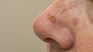
A rough, scaly or sandpaper-like spot on your face, scalp, ears, forearms or hands may be an actinic keratosis. Often called an AK, this common precancerous growth develops after years of exposure to ultraviolet light from the sun or indoor tanning. It is one of the most common reasons people see a dermatologist, and in a region where so much of life happens on the water, on a flight line, or on a golf course, we see a great deal of it.
An estimated 58 million Americans have at least one actinic keratosis, according to a national report on the burden of skin disease cited by the Skin Cancer Foundation. Most people who develop an AK will develop more. The good news is that dermatologists can successfully clear many actinic keratoses when they find and treat them early, reducing the risk that these lesions will progress to skin cancer.
What is actinic keratosis?
An actinic keratosis is a small area where the cells in the top layer of your skin start growing abnormally. The cause is cumulative ultraviolet exposure, either from the sun or from indoor tanning. “Actinic” simply means caused by radiation, which is why you may also hear these spots called solar keratoses.
Dermatologists consider actinic keratoses precancerous. They are not skin cancer, but dermatologists consider them precancerous because some can develop into squamous cell carcinoma. Aks are a reliable signal that the surrounding skin has taken on real sun damage as well. When you have several in one area, such as a balding scalp or a forearm, we describe that area as a field of actinic damage. That distinction matters a great deal when it comes to choosing treatment.
How to pronounce actinic keratosis
Actinic keratosis is pronounced ak-TIN-ik ker-uh-TOH-sis. More than one is called actinic keratoses, pronounced ker-uh-TOH-seez. Your dermatologist might refer to it as an “AK”.
What Actinic Keratosis Looks and Feels Like
Most people feel an actinic keratosis before they see it. The classic description is a patch of sandpaper on otherwise smooth skin, often noticed while shaving, washing your face, or drying off. Individual spots range from smaller than a pencil eraser to about an inch across, and they can be flat or slightly raised.
Rough, scaly patches
The most common presentation is a dry, gritty patch that catches on a towel or a razor. It may flake, and it may be easier to locate with your fingertips than with a mirror.
Red or pink raised bumps
Many actinic keratoses appear as small red or pink bumps with a scaly surface. They tend to cluster on the areas that catch the most sun over a lifetime: the nose, cheeks, ears, upper lip, bald scalp, neck, forearms, and hands.
Brown or tan patches
These can be difficult to tell apart from age spots, seborrheicSome actinic keratoses contain pigment and appear as flat brown or tan patches. keratoses, or in some cases melanoma, which is one of the better reasons to have a new brown patch evaluated.
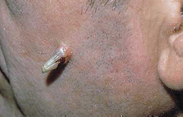
Thick Horn-Like Growths
Occasionally an actinic keratosis builds up dense keratin and grows outward into a hard projection, sometimes called a cutaneous horn. Dermatologists call the thicker variety hypertrophic actinic keratosis. These are more likely to harbor cancer cells and generally need a biopsy rather than simple freezing.
Actinic cheilitis on the lips
When sun damage affects the lips—usually the lower lip—dermatologists call it actinic cheilitis. Instead of a discrete patch, the lip becomes persistently dry, rough, and chapped in a way that lip balm never quite resolves, sometimes with a blurred border between the lip and the skin above it. It is easy to dismiss for years. It deserves the same attention as an actinic keratosis anywhere else.
Actinic keratosis on darker skin tones
Actinic keratoses most commonly affect people with fair skin, but they can also develop in people with brown and Black skin, where they may be harder to detect. On deeper skin tones the redness that makes a lesion obvious on fair skin may be subtle or absent, and the spot may read as a grayish, hyperpigmented, or simply rough area. If you have a patch of skin that feels different from the skin around it, texture is the more reliable clue than color.
Concerned about a rough or scaly spot?
Our board-certified dermatologists diagnose and treat actinic keratoses at eight convenient Hampton Roads locations.
Can actinic keratosis come and go?
Yes, and this is one of the most commonly misunderstood things about the condition. An actinic keratosis can appear, stay for weeks or months, flake away, and leave skin that feels perfectly smooth. Then it comes back, often after a weekend outdoors without sun protection.
This is well documented. Published reviews report that roughly a quarter of individual actinic keratoses regress on their own within about a year. The catch is that a meaningful share of those, approximately 15 percent, return afterward. Spontaneous disappearance is real, and is not the same thing as resolution.
The reason is straightforward. What flakes off is the visible surface. The underlying ultraviolet damage to the DNA in those skin cells is still there, and so are the neighboring cells that took the same damage without yet producing a visible spot. A spot that comes and goes is telling you that the skin in that area is unstable, not that the problem has passed. It is worth an examination even if it is not there on the day of your appointment, because your dermatologist can assess the whole field.
Is actinic keratosis skin cancer?
No. An actinic keratosis is a precancer, which means it is not cancer but can develop into cancer if it is left alone. The specific concern is squamous cell carcinoma, the second most common form of skin cancer.
The numbers help put this in perspective. The chance that any individual actinic keratosis will become squamous cell carcinoma is difficult to predict and estimates vary. Because dermatologists cannot reliably determine which lesions will progress, treatment is usually recommended. A 2021 study in JAMA Dermatology found that just under 9 percent of patients with actinic keratoses had developed a squamous cell carcinoma at roughly four years of follow-up. Looked at from the other direction, published research indicates that a large majority of squamous cell carcinomas arise from pre-existing actinic keratoses.
The risk from any one spot is low, and the risk across many spots over many years is higher. The practical problem is that there is currently no way to look at a given actinic keratosis and know whether it is one of the ones that will progress. That is why dermatologists treat them rather than monitor them. Caught at the precancer stage, treatment takes minutes. Caught later as a squamous cell carcinoma, it may require excision or Mohs surgery.
What causes actinic keratosis, and who is most at risk?
Actinic damage accumulates. The exposure that produces an actinic keratosis in your sixties was largely banked decades earlier, which is why these spots show up on the areas you have never been able to keep covered. Risk is higher if you:
- Are over 45, with risk continuing to climb with age
- Have fair skin, light eyes, or blond or red hair, and burn before you tan
- Have worked or played outdoors for years, particularly more than a few hours a day
- Have used tanning beds
- Have a bald or thinning scalp
- Have had a skin cancer or an actinic keratosis before
- Take a medication that increases sun sensitivity
- Have a weakened immune system, whether from a medication, a condition, or an organ transplant
That last group deserves emphasis. Organ transplant recipients on long-term immunosuppression carry a substantially elevated lifetime risk of actinic keratoses and of skin cancer, and they generally need closer surveillance than the general population.
Age alone accounts for a great deal. A 2022 JAMA Dermatology analysis of a large group of Medicare patients aged 65 and older found that nearly 30 percent had been diagnosed with at least one actinic keratosis.
Schedule An Appointment
Why Hampton Roads Sees So Much Sun Damage
This region has a large concentration of people whose lives involve sustained outdoor ultraviolet exposure. Hampton Roads supports 18 military installations and roughly 120,000 active-duty, reserve, and civilian defense personnel, along with more than 125,000 military dependents and close to 220,000 veterans, according to the Hampton Roads Military and Federal Facilities Alliance. Naval Station Norfolk is the largest naval base in the world.
Years of deck watches, flight-line work, shipyard shifts, and field training add up to a career of exposure that no weekend of sunscreen offsets. The same is true for the region’s watermen and commercial fishermen working the Chesapeake Bay and the more than 3,000 miles of shoreline the Coast Guard’s Sector Virginia covers, and for anyone who has spent decades at the oceanfront, on a boat, or on a golf course.
Water and wet sand reflect ultraviolet light back at you, so a day on the Bay or at the beach delivers considerably more than the same day inland. And ultraviolet light passes through cloud cover, which means the overcast, humid days are not the “safe days” they feel like. If you spent your twenties and thirties working outdoors, actinic keratoses in your fifties and sixties are not a surprise, and they are worth examining.
How Dermatologists Diagnose Actinic Keratosis
Most actinic keratoses are diagnosed on examination. Your dermatologist will look at the spot and the skin around it, and will usually feel it, because texture carries real diagnostic information. A dermatoscope, a handheld lens with polarized light, helps clarify the surface pattern and distinguish an actinic keratosis from other lesions that resemble it.
A biopsy is recommended when a spot is thick, tender, bleeding, ulcerated, growing quickly, or simply not behaving the way an actinic keratosis should. This is not excessive caution. Studies have found that a meaningful percentage of lesions diagnosed clinically as actinic keratoses turn out on pathology to be squamous cell carcinoma, and there is no way to catch that without looking. A biopsy is a brief in-office procedure done with local anesthetic. Pariser Dermatology sends specimens to our own in-house dermatopathology lab at our Norfolk office, where board-certified dermatopathologists read them, which can mean a faster answer than practices that ship tissue to an outside laboratory. This also allows our dermatologists to collaborate with our pathology specialists.
Actinic Keratosis Treatment Options
Treatment falls into two categories, and understanding the difference explains most of what your dermatologist will recommend. Lesion-directed treatment removes the spots you can see. Field therapy treats an entire area of sun-damaged skin, including the abnormal cells that have not yet surfaced. Many patients need both, sometimes in sequence.
In-office treatments for individual spots
- Liquid nitrogen is applied to the spot for a few seconds, freezing and destroying the abnormal cells. This is the most common treatment for actinic keratosis. The treated area typically stings briefly, then blisters or scabs and peels within one to three weeks. It sometimes leaves a small pale mark.
- Curettage, sometimes with electrosurgery. The lesion is scraped away with a curette, a small spoon-shaped instrument, and the base may be treated with electrosurgery. This has the advantage of producing tissue that can be sent for pathology, which is useful for thicker lesions.
- Medical chemical peel. A prescription-strength peel applied in the office removes the damaged upper layers of skin so healthy skin can replace them. This is a medical procedure and is not the same as anything available at a spa or in an at-home kit.
- Laser resurfacing: Much like a chemical peel, a laser can remove the surface layer of the skin. This destroys AK cells. After treatment, the skin will be raw and sore. The skin heals within 1 or 2 weeks, revealing healthier new skin.
Field therapy for widespread sun damage
If you have many actinic keratoses across a scalp, forehead, or forearm, freezing them one at a time addresses only what is visible and leaves the rest of the damaged field in place. Field therapy treats the whole area.
- 5-fluorouracil (5-FU) cream. A topical chemotherapy applied at home, typically twice daily for two to four weeks. It works by making the abnormal cells react, so the treated skin becomes red, raw, and crusted before it heals. That reaction is expected and is a sign the medication is doing its job, though it is worth planning around socially.
- Imiquimod cream. Rather than destroying cells directly, imiquimod prompts your immune system to clear them. It is applied on a schedule your dermatologist sets, usually over several weeks, and also produces redness and inflammation.
- Tirbanibulin (Klisyri) ointment. The newest option and by far the shortest course: once daily for five consecutive days. It was FDA approved in December 2020, and in June 2024 the approved treatment area was expanded fourfold, to fields of up to 100 square centimeters on the face or balding scalp. In two phase 3 trials involving 702 adults, complete clearance of lesions in the treated area was achieved in 44 and 54 percent of patients, compared with 5 and 13 percent on placebo. Local redness and flaking are common but the reaction is generally shorter-lived than with older topicals, which makes it a practical choice for patients who cannot commit to weeks of visible irritation.
- Diclofenac sodium gel. A gentler, slower option applied twice daily for roughly two to three months. It causes less dramatic inflammation, which suits some patients, but it requires patience and diligent sun protection throughout.
- Photodynamic therapy. A light-sensitizing solution is applied to the treatment area and left to absorb, then activated with a specific wavelength of light in the office. The activated solution destroys the abnormal cells. Photodynamic therapy treats a broad field and is often chosen for scalps and faces with extensive damage. Expect redness and peeling for several days, and strict sun avoidance immediately afterward.
A note about Picato
You may come across older articles, and even some practice websites, recommending Picato (ingenol mebutate) as a two-day or three-day gel treatment for actinic keratosis. It is no longer available. The manufacturer withdrew Picato from the market in 2020 after studies linked it to a higher rate of skin cancer—particularly squamous cell carcinoma—compared with imiquimod. European regulators concluded that its risks outweighed its benefits. If you used Picato in the past, tell your dermatologist at your next skin check so they can carefully examine the treated areas.
Schedule An Appointment
What to Expect after Treatment
Healing depends on the treatment method. Cryotherapy and curettage usually create a scab that resolves in one to three weeks. Field treatments take longer, because the skin has to react before it can clear, and most patients see redness, flaking, and tenderness for one to several weeks depending on the medication.
Sun protection during and after treatment is mandatory. Treated skin is temporarily more vulnerable to ultraviolet light, and new exposure works directly against what the treatment is trying to accomplish.
Follow-up depends on how much sun damage you have. Patients with a heavy burden of actinic keratoses should return every 8 to 12 weeks, at least initially. Patients with a few isolated spots may only need an annual skin cancer screening. Either way, keeping those appointments is the point. Surveillance is what turns a lifetime of sun damage into a manageable condition.
Why Actinic Keratosis Can Come Back after Freezing
This is one of the most frequent questions we hear, and recurrence after cryotherapy is not indicative of a failed treatment. There are a few reasons it happens.
- Freezing treats the visible lesion. The treatment leaves the surrounding sun-damaged cells untouched, allowing new spots to develop.
- Thicker, hypertrophic actinic keratoses can be difficult to freeze all the way through in one pass, and may need a second treatment or a different approach entirely.
- The original cause has not gone away. The ultraviolet damage banked over decades is permanent, and continued exposure keeps generating new lesions.
If spots keep returning in the same area, that is usually a signal to move from lesion-directed treatment to field therapy, or to combine the two. A frequent and effective approach is to freeze the thickest lesions first and then treat the surrounding field with a topical or with photodynamic therapy.
Actinic Keratosis Compared with Other Skin Spots
Actinic keratosis vs. seborrheic keratosis
The names are similar and the conditions are not related. A seborrheic keratosis is a harmless growth that tends to look waxy or warty and appears stuck onto the skin, often light tan darkening to brown or black over time. It has no relationship to skin cancer. An actinic keratosis is flatter, rougher, drier, usually pink or red, and it is precancerous. If you cannot tell which you are looking at, that is entirely reasonable, and it is worth an appointment.
Actinic keratosis vs. dry skin or eczema
Dry skin and eczema respond to moisturizer, typically improve within days, and usually affect broader areas symmetrically. An actinic keratosis is a discrete, persistent patch in a sun-exposed location. It does not improve with moisturizer no matter how diligently you apply it. A rough spot that has survived a few weeks of good skin care is not dry skin.
Actinic keratosis vs. squamous cell carcinoma
Because one can become the other, the distinction can be subtle. Signs that a spot has progressed include noticeable thickening or firmness, tenderness or pain, bleeding, an open sore that will not heal, or rapid growth. Any of those warrants prompt evaluation and probably a biopsy.
How to Lower Your Risk of New Actinic Keratoses
You cannot undo existing sun damage, but you can substantially slow the rate at which new actinic keratoses form. The habits that matter most:
- Apply broad-spectrum sunscreen of SPF 30 or higher every day to your face, ears, neck, and hands, and reapply every two hours outdoors
- Wear a wide-brimmed hat, which protects the scalp, ears, and neck far better than a ball cap
- Choose sun-protective clothing with a UPF rating for long days on the water or on the job
- Seek shade during the strongest hours, roughly 10 a.m. to 4 p.m.
- Skip tanning beds entirely
- Check your own skin paying attention to texture as well as appearance
Talk with a Hampton Roads Dermatologist about that Rough Patch
Pariser Dermatology Specialists has been caring for Hampton Roads skin since 1946, and we remain independent and locally owned. In practical terms, your dermatologist determines your treatment plan without pressure from an outside owner. Some of our physicians teach at Eastern Virginia Medical School, and our dermatopathologists read biopsies in our own Norfolk lab. Through Virginia Clinical Research our patients have access to studies of emerging treatments for sun damage and skin cancer.
If you have a rough or scaly spot that has not cleared on its own, or you have not had your skin checked in over a year, our team is glad to take a look. To schedule an appointment with one of our board-certified dermatologists at any of our eight Hampton Roads locations, call us at 757-622-6315 or schedule your appointment online.
Schedule An Appointment